Small nanoparticles were already being used by the Romans to color glass. When applied in the right way, small traces of gold, silver and copper can create intensively colored glass. This color is not only produced by the selective absorption of specific wavelengths like in normal dyes but more so by extensive scattering of light with the resonance wavelength of the plasmon in these nanoparticles. This effect is also responsible for the blue color of the sky and the red color at dawn. Small particles, or inhomogeneities, of the atmosphere scatter the yellow sunlight most effectively in the blue spectral range which makes the sky look blue during the day. At dawn, the light has to travel such a long distance in the scattering atmosphere, resulting in most of the blue light being dispersed, leaving only the complementary color red. The color of the famous Lycurgus cup, which is exhibited at the British Museum in London, can be explained in a similar way. Depending on whether the light source is in front of or behind the cup, the color appears to be red or green.
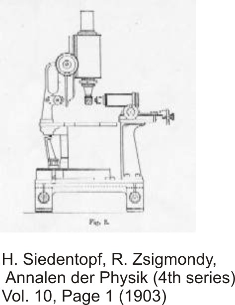
 Lord Faraday was the first to scientifically examine the color of small gold particles, and he gained some impressive insight. A good 30 years later, Richard Zsigmondy also extensively studied the properties of nanoparticles and was awarded the Nobel Prize in Chemistry in 1925 partly for his work on this topic. He developed ultra microscopy, which is called dark field microscopy today, in cooperation with Siedenkopf. Lacking the possibilities to analyze the spectra as well as the shape and size of single particles through electron microscopy caused many of their experiments to be only of a qualitative nature.
 On the right is possibly the first color picture showing light scattering in single gold particles, published by Zsigmondy in 1907 in Jena. The picture is not a photograph but an artistic depiction! However, the colors resemble the real colors in sunlight very well. If you compare this picture to the ones in the research section on this homepage, you will see that although the techniques are a lot more advanced these days, the images look very much alike.
Dark field microscopy was partly replaced by fluorescence microscopy but in recent years, regained importance due to the growing interest in nano structures. A good, comprehensive introduction to dark field microscopy can be found here.
